.svg)
Understanding Dog Bite Scarring: Types and Anatomy of Canine Wounds
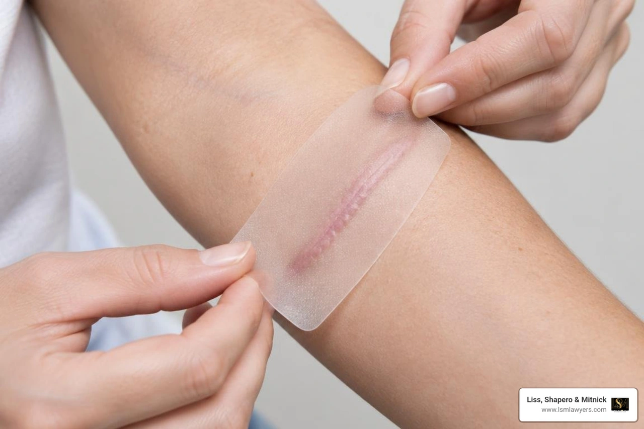
Dog bite scarring refers to the permanent marks left after a canine bite heals. These scars form when the skin's deep layers are damaged by crushing, puncturing, or tearing forces, and they often appear raised, discolored, or irregular. Treatment focuses on minimizing scar visibility through early wound care, silicone therapy, sun protection, and sometimes surgical revision. Healing typically takes 12 to 18 months, during which the scar gradually softens and fades.
Canine jaws exert incredible pressure, often exceeding 200 to 400 pounds per square inch depending on the animal's size and breed. Unlike the clean, sharp cut of a surgical scalpel or kitchen knife, canine trauma combines crushing pressure, puncture forces, and mechanical tearing. This mechanism causes devitalized tissue margins, subcutaneous bruising, and deep structural damage that severely complicates healing.
Furthermore, a dog’s oral cavity harbors virulent polymicrobial flora, including Pasteurella multocida, Staphylococcus aureus, Streptococcus, and Capnocytophaga canimorsus. When teeth puncture the dermis, they seed these bacteria into deep, poorly oxygenated tissue spaces. The ensuing bacterial proliferation triggers acute inflammation, which breaks down healthy extracellular matrix and degrades local collagen networks.
When tissue viability is compromised and bacterial loads remain high, the body repairs the defect by laying down haphazard, disorganized collagen bundles rather than smoothly organized dermal layers. For a detailed breakdown of these trauma patterns, read our comprehensive 2026 dog bite guide.
Punctures, Lacerations, and Avulsion Defects
Dog bite wounds generally fall into three distinct anatomical categories:
- Puncture Wounds: Narrow, deep wounds caused by the canine teeth. Punctures often look deceptively benign on the surface, but they inject bacteria deep into subcutaneous fat, fascia, and tendon sheaths. Because they heal from the outside in, trapped bacteria frequently cause localized abscesses that worsen internal scarring.
- Jagged Lacerations: Created when a dog bites and pulls, or when a victim pulls away. The shearing force leaves irregular, ragged wound margins with compromised microcirculation. These devitalized edges frequently undergo micro-necrosis, leading to widened, uneven scars.
- Avulsion Defects: The most severe bite pattern, occurring when skin, subcutaneous tissue, and underlying muscle are torn entirely away from the body. Because the wound edges cannot be pulled together directly, these injuries require complex grafting, local flaps, or prolonged secondary-intention healing, all of which leave extensive, thick scars.
How Location and Age Influence Dog Bite Scarring
Anatomical location directly shapes the aesthetic and functional outcome of a bite wound. Wounds situated across natural relaxed skin tension lines (Langer’s lines)—such as across the forehead, perpendicular to the jawline, or over joint surfaces—experience constant mechanical pulling during normal movement. This chronic tension stimulates fibroblasts to overproduce collagen, creating widened, raised, or contractured scars that restrict physical mobility.
Facial trauma represents one of the most critical challenges. The lips, nose, eyelids, and ears possess delicate micro-architecture. Lacerations crossing the vermilion border of the lip, for instance, demand sub-millimeter surgical realignment. Even a one-millimeter step-off deformity creates a visibly glaring irregularity that requires future scar revision.
Age also plays a crucial role in wound behavior:
- Children: Children under five suffer facial bites at disproportionately high rates due to their height relative to medium and large dogs. Pediatric skin exhibits high elasticity and rapid cellular turnover, which supports rapid wound closure. However, robust pediatric immune responses frequently generate hyperactive, erythematous, and hypertrophic scars.
- Adults: Adult victims sustain the majority of dog bites to the extremities, particularly the hands and forearms. Adult skin possesses lower collagen turnover and reduced vascular elasticity, increasing the risk of delayed healing, wound breakdown, and stiff, atrophic scarring across finger joints and wrists.
Immediate Wound Care and the Clinical Healing Timeline
The final cosmetic appearance of a canine bite depends heavily on the care delivered within the first 60 minutes of injury. Proper emergency hygiene dramatically curtails bacterial loads, halting infectious destruction before deeper tissue is lost.
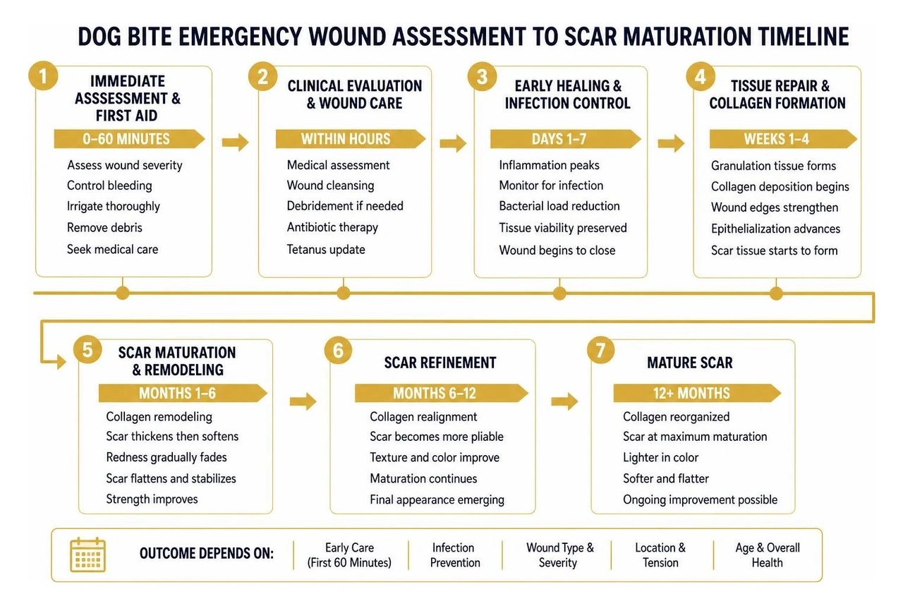
A structured approach to recovery minimizes complications. For an overview of this multi-stage healing journey, review the dog bite wound care timeline.
First-Aid Protocol and Infection Prevention
If you or a loved one sustains a dog bite, follow this clinical first-aid protocol immediately:
- Hemostasis: Apply continuous, firm pressure with a clean cloth or sterile gauze for 5 to 10 minutes to stop active bleeding.
- Copious Irrigation: Flush the wound thoroughly with lukewarm running tap water or sterile normal saline for at least 5 to 10 minutes. Avoid harsh antiseptics like undiluted hydrogen peroxide, rubbing alcohol, or povidone-iodine inside the wound bed, as they kill delicate cellular membranes and delay re-epithelialization.
- Gentle Cleansing: Wash the surrounding intact skin gently using mild unscented soap, keeping soap out of deep open punctures to prevent chemical irritation.
- Sterile Dressing: Apply a thin layer of over-the-counter bacitracin or petrolatum ointment and cover the injury with a sterile, non-adherent dressing.
- Urgent Medical Care: Seek professional evaluation at an urgent care or emergency room within 8 hours. Clinicians will assess the need for prophylactic oral antibiotics (typically amoxicillin-clavulanate), a tetanus booster (if your last dose was over 5 years ago), and rabies post-exposure prophylaxis.
For practical Michigan legal and medical guidance right after an attack, consult our resource on immediate steps after a Michigan dog bite.
The 12-Month Scar Maturation Process
Scar maturation is not a single event—it is a continuous biological process spanning roughly a full calendar year:
- Hemostasis and Inflammatory Phase (Days 0 to 7): Platelets aggregate, forming a fibrin clot. White blood cells migrate to the injury site to clear cellular debris and destroy bacteria. The wound exhibits redness, localized warmth, and swelling.
- Proliferative Phase (Weeks 1 to 6): Fibroblasts flood the area, generating a provisional matrix of disorganized Type III collagen. New capillary buds form via angiogenesis. The newly closed scar typically appears bright red, rigid, and slightly raised.
- Remodeling and Maturation Phase (Months 2 to 12+): The body gradually replaces fragile Type III collagen with stronger, organized Type I collagen. Blood vessels regress, causing the initial redness to fade to pink and eventually to a pale, flesh-toned hue. The scar's tensile strength plateaus at approximately 70% to 80% of uninjured skin.
Plastic surgeons typically advise waiting the full 12 months before considering definitive surgical scar revision, ensuring the tissue has reached cellular maturity.
Non-Surgical and Surgical Scar Management Strategies
Reconstructive surgeons evaluate bite injuries using the "reconstructive ladder," starting with non-invasive therapies before escalating to complex surgical techniques. For complex facial cases involving tissue loss, see this clinical pediatric lower-lip reconstruction study.
Non-Surgical Treatments for Dog Bite Scarring
Conservative scar therapies should begin as soon as the epidermal barrier is intact and all sutures have been removed:
- Silicone Therapy (Sheets and Gels): Medical-grade silicone remains the gold standard in non-invasive scar management. Silicone hydrates the stratum corneum, normalizes fibroblast signaling, and reduces collagen overproduction. Silicone sheets or gels should be worn 12 to 24 hours daily for 3 to 6 consecutive months.
- Targeted Pressure Garments: Custom elastic pressure garments apply continuous mechanical compression (20–30 mmHg) to scars on the extremities. This limits local tissue perfusion and inhibits excessive fibroblast activity.
- Intralesional Corticosteroid Injections: Triamcinolone acetonide (10–40 mg/mL) injected directly into raised, rigid scars breaks down excessive collagen bonds, reduces pruritus, and flattens thickened tissue.
- Vascular and Fractional Laser Therapy: Pulsed-dye lasers (PDL, 585–595 nm) target hemoglobin in micro-vessels to eliminate persistent scar erythema. Fractional ablative (CO2) and non-ablative lasers create microscopic thermal treatment zones, stimulating controlled dermal remodeling to blend jagged scar margins with adjacent skin.
- Medical Microneedling: Controlled mechanical micro-punctures stimulate localized collagen restructuring, softening rigid scar margins and improving skin texture.
- Strict Photoprotection: Healing scar tissue lacks mature melanin regulation. Ultraviolet exposure triggers profound post-inflammatory hyperpigmentation that can become permanent. Apply a broad-spectrum, mineral-based (zinc oxide/titanium dioxide) sunscreen with SPF 30+ daily for at least 12 to 18 months.
Surgical Scar Revision and Reconstructive Procedures
When conservative modalities cannot correct substantial step-off deformities, webbing, or tissue distortion, reconstructive surgery becomes necessary.
| Procedure | Primary Indication | Clinical Approach | Expected Outcome |
|---|---|---|---|
| Surgical Excision & Layered Closure | Wide, jagged, or hyper-pigmented scars | The irregular scar is excised cleanly, and subcutaneous layers are mobilized and sutured in tension-free anatomical layers. | A thin, flat, linear scar that aligns with natural skin tension lines. |
| Z-Plasty / W-Plasty | Webbed scars, joint contractures, or scars crossing tension lines | Triangular tissue flaps are transposed to break up linear contractures and change scar direction by up to 90 degrees. | Relieves mechanical tension, restores full joint mobility, and camouflages straight scar lines. |
| Composite Grafting | Severe avulsion defects involving skin, fat, and cartilage/mucosa | Multilayered tissue (often harvested from donor sites or salvaged tissue) is grafted into the defect. | Restores complex anatomical contours, such as the nasal rim or lip vermilion. |
| Full-Thickness Skin Grafts (FTSG) | Extensive surface skin loss where primary closure causes distortion | Complete epidermal and dermal layers are harvested (e.g., from the supraclavicular area or groin) and secured over the wound bed. | Prevents graft contracture and provides durable, color-matched coverage. |
| Tissue Expansion | Substantial tissue loss requiring matching local skin | An inflatable silicone balloon is placed beneath adjacent healthy skin and filled gradually with saline over several weeks. | Generates excess matching skin to cover the defect with identical color, texture, and hair-bearing qualities. |
Managing Scars in Highly Melanated Skin: Keloid and Hypertrophic Risks
Patients with Fitzpatrick skin phototypes IV through VI (including individuals of African, Hispanic, Middle Eastern, and Asian descent) possess melanocytes that respond aggressively to cutaneous trauma. About 4.5% to 16% of the general population develops abnormal scarring, with a substantially higher frequency observed in highly pigmented skin (Sheth & Thakur, 2024).
For deep insights into the surgical complexities of managing severe canine trauma in darker skin types, refer to this Cureus case report on avulsion dog bites.
Keloid vs. Hypertrophic Scar Pathophysiology
Distinguishing between hypertrophic scars and true keloids is essential for designing an effective treatment strategy:
- Hypertrophic Scars: Raised, erythematous, rigid scars that remain strictly confined within the boundaries of the original dog bite laceration. They typically develop within 4 to 8 weeks post-injury, undergo a rapid growth phase, and may slowly regress over several years.
- Keloid Scars: Benign dermal fibroproliferative tumors composed of dense, hyalinized collagen bundles that expand well beyond the margins of the original wound, invading surrounding healthy tissue. Keloids rarely regress on their own and frequently recur after surgical excision.
Specialized Clinical Protocols for Darker Skin Tones
Treating dog bite wounds in patients with darker skin requires specialized surgical and dermatological techniques to prevent excessive collagen formation and severe dyschromia:
- Minimal Incision & Low-Tension Techniques: Surgeons minimize the number of auxiliary incisions and avoid aggressive tissue undermining. Multi-layered dermal suspension sutures are used to eliminate wound-edge tension.
- Salvaged Tissue Composite Grafting: In severe avulsion bites, plastic surgeons may clean, debride, and reuse the patient's own avulsed tissue as a composite graft, eliminating the need for a secondary donor site that could develop a new keloid (Sheth & Thakur, 2024).
- Combined Intralesional Steroids & 5-Fluorouracil (5-FU): Blending triamcinolone acetonide with 5-FU limits fibroblast proliferation while reducing steroid-induced subcutaneous fat atrophy and telangiectasia.
- Conservative Cryotherapy: Controlled intralesional cryoneedle application freezes the scar from the inside out, preserving surface melanocytes to avoid permanent skin depigmentation.
- Post-Operative Superficial Radiation Therapy (SRT): For severe, recurring keloids resistant to other therapies, low-dose superficial radiation delivered within 24 to 48 hours of surgical excision can inhibit fibroblast mitosis and prevent regrowth.
Psychological Trauma and Legal Compensation for Disfigurement
The physical marks left by a canine attack represent only a portion of the total injury. The sudden, violent nature of an animal attack can cause profound, enduring psychological harm.
Emotional Recovery and Long-Term Mental Health
Between 25% and 30% of dog bite victims develop clinically significant Post-Traumatic Stress Disorder (PTSD). Symptoms frequently include:
- Cynophobia: An acute, debilitating fear of dogs that disrupts daily activities, outdoor exercise, and social interactions.
- Intrusive Flashbacks & Sleep Disturbances: Recurrent nightmares and intrusive memories of the attack triggered by barking or animal sightings.
- Body Dysmorphic Distress: Severe social anxiety, depression, and lowered self-esteem stemming from visible facial or bodily disfigurement.
- Pediatric Behavioral Regression: Children may experience bedwetting, separation anxiety, panic attacks, and severe social withdrawal due to playground teasing or bullying.
Professional psychological interventions—such as trauma-focused Cognitive Behavioral Therapy (CBT) and Eye Movement Desensitization and Reprocessing (EMDR)—are often essential components of comprehensive medical recovery.
Documenting Scars to Maximize Legal Recovery
Under Michigan law (MCL 287.351), dog owners face strict liability for bite injuries when the victim was lawfully on the property and did not provoke the dog. In addition to medical bills and lost wages, victims are entitled to pursue non-economic damages for physical pain, emotional distress, and permanent scarring or disfigurement.
Because insurance carriers frequently try to settle claims early before the full extent of scarring is known, meticulous documentation is critical:
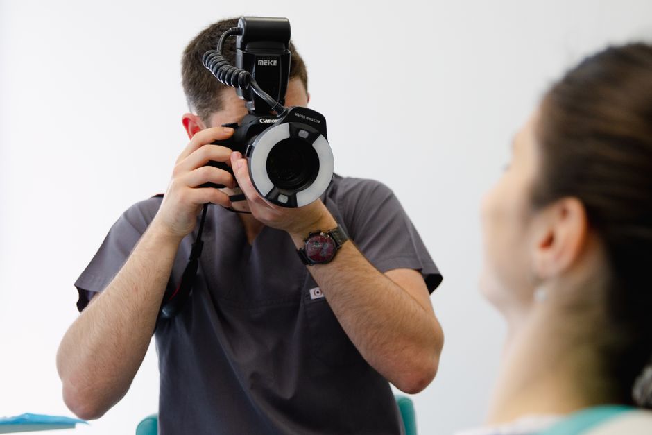
- High-Resolution Serial Photography: Capture clear, well-lit, non-flash photographs of the wound immediately after the bite, throughout the suturing and recovery phases, and at 3, 6, 9, and 12 months. Include close-up shots alongside wider framing to show the scar's position relative to your entire face or limb.
- Independent Medical Evaluations: Consult a board-certified plastic surgeon to obtain formal scar evaluations and detailed cost projections for future revisions, laser therapies, or reconstructive procedures.
- Comprehensive Mental Health Records: Document all therapy visits, clinical psychological assessments, and prescription medications used to treat attack-related trauma.
Understanding how insurance carriers handle these injuries is critical; learn more about navigating a homeowner's insurance liability for dog bites claim. For an in-depth breakdown of non-economic damage calculations, review our guide on evaluating dog bite claim value.
Frequently Asked Questions About Dog Bite Scars
How long does a dog bite scar take to fully mature?
A dog bite scar takes between 12 and 18 months to fully mature. During the first 6 to 12 weeks, scars often appear red, raised, and firm due to heightened vascularity and active collagen deposition. Over the subsequent 9 to 12 months, the tissue remodels, blood vessels regress, and the scar softens and fades. Reconstructive surgeons generally advise waiting at least one full year before undergoing elective surgical scar revision.
Can laser therapy or surgery completely remove a dog bite scar?
No medical procedure can completely erase a scar or restore skin to its original, uninjured state. However, modern treatments can dramatically reduce scar visibility. Vascular lasers can eliminate persistent redness, fractional CO2 lasers can smooth irregular surface textures, and surgical revisions (such as Z-plasties) can realign scars along natural skin creases, making them far less noticeable.
What factors determine compensation for permanent dog bite disfigurement?
Compensation for permanent scarring depends on several key legal and medical factors:
- Anatomical Location: Scars on prominent facial regions (lips, cheeks, nose) or the hands command substantially higher compensation than scars easily hidden by clothing.
- Age and Gender of the Victim: Disfigurement in young children frequently results in higher damages due to the lifelong social and psychological burden they carry.
- Severity and Type of Scarring: Thick hypertrophic scars, large keloids, or contractures that limit joint movement increase non-economic claim value.
- Projected Medical Costs: Formal estimates from plastic surgeons detailing the necessity and cost of future revisions, laser treatments, or skin grafting.
- Available Insurance Coverage: Homeowner and renter insurance liability policy limits carried by the dog's owner.
Conclusion
Recovering from a severe dog bite requires a comprehensive, two-fold approach: thorough medical care to optimize physical healing and assertive legal representation to secure the resources needed for long-term recovery. Scar prevention begins with immediate wound cleaning and infection control, followed by long-term scar care using silicone therapy, sun protection, and specialist-guided reconstructive procedures.
At Liss, Shapero & Mitnick, our legal team draws on decades of experience—including valuable insight gained from our background in insurance defense—to advocate effectively for dog bite victims across Berkley, Metro Detroit, and all of Michigan. We know the tactics insurance adjusters use to downplay long-term disfigurement, and we work diligently to ensure your settlement covers all current and future medical needs, reconstructive surgeries, and emotional trauma.
If you or a family member is dealing with lasting scars from an animal attack, explore our guide on documenting dog bite scars and claim value drivers to protect your health and legal rights today.
References
Sheth R, Thakur M. "The Surgical Repair of an Avulsion Dog Bite and Scar Revision in Highly Melanated Skin: A Case Report.." Cureus, 2024. PMCID PMC11668903.