
How to Judge Dog Bite Scar Severity Before You Settle
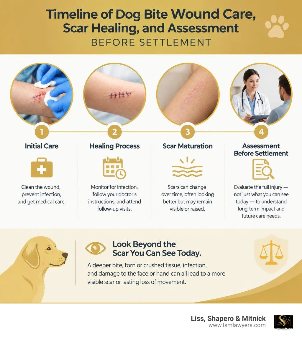
Look at more than the scar you can see today. A deeper bite, torn or crushed tissue, infection, and damage to the face or hand can all lead to a more visible scar or lasting loss of movement. Take clear photos, keep your treatment records, and ask your doctor whether you may need scar treatment or surgery. Do not judge a settlement offer by how the wound looks while it is still healing.
I’m Anthony Shapero, Managing Partner at Liss, Shapero & Mitnick. My years representing injured people and my earlier work in insurance defense help me assess how insurers may view dog bite scar injury severity, future care, and the cost of permanent disfigurement.
Dog bite scar injury severity helpful reading:
Clinical Determinants of Dog Bite Scar Injury Severity
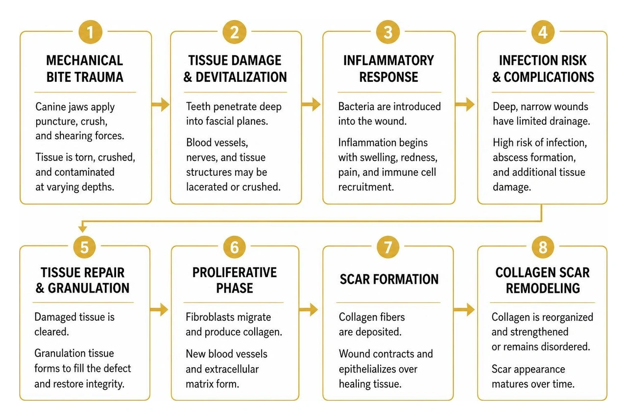
Animal bites account for approximately 337,000 emergency department visits across the United States every year. When an animal attacks, the resulting wound is rarely a clean, surgical incision. Canine dentition exerts massive mechanical force—often between 200 and 400 pounds per square inch depending on the animal's size—delivering a complex combination of puncture, crush, and shearing forces.
Evaluating the clinical determinants of dog bite scar injury severity requires understanding identifying what a dog bite looks like across wound types and how specific trauma mechanisms compromise tissue viability. The ultimate cosmetic outcome depends on the degree of tissue devitalization, structural damage, underlying vascular injury, and the body's inflammatory response during wound remodeling.
How Wound Depth and Mechanism Shape Dog Bite Scar Injury Severity
Canine jaws produce three distinct wound mechanisms, often simultaneously:
- Puncture Wounds: Canine teeth penetrate deep into underlying fascial planes, inoculating bacteria while leaving a deceptively small surface entry point. Because these wounds lack natural drainage, they harbor high risks of closed-space infection and deep subcutaneous fibrosis.
- Crush Trauma: The compressive force of the jaws devitalizes skin margins, bruises subcutaneous fat, and compromises microvascular perfusion. Devitalized margins cannot heal by primary intention without surgical debridement, significantly increasing the risk of broad, dense scarring.
- Avulsion and Shearing Lacerations: When a dog bites and pulls, or when a victim pulls away, shearing forces tear skin and subcutaneous fat away from underlying fascia. This causes soft tissue avulsion flaps, disrupting blood supply and leaving irregular wound edges that heal with uneven, hypertrophic margins.
Clinical studies on child dog bite injuries reveal that the majority of pediatric patients sustain deep tissue trauma involving structural layers rather than simple abrasions. When muscle fascia or periosteum is violated, fibrotic remodeling extends through multiple tissue planes, creating anchored, tethered scars that restrict natural movement and distort resting skin contour.
Anatomical Site Vulnerability and Cosmetic Impact
The location of a dog bite strongly dictates both the biological risk of infection and the cosmetic impact of the resulting scar. Canine bites to the face heal with different clinical dynamics than bites to the extremities due to variations in vascularity, skin tension, and underlying anatomy.
- Head and Neck: The face possesses an exceptionally rich vascular supply, which supports rapid tissue survival and lowers baseline infection rates to between 4% and 11%. However, because the face is the focal point of human communication and identity, even minor contour irregularities, hyperpigmentation, or resting tension line mismatches produce severe cosmetic disfigurement. Injuries across the vermilion border of the lips, nasal alar base, or eyelid margins require microscopic alignment to prevent permanent structural distortion.
- Hands and Wrists: The hands present the highest biological risk, with infection rates ranging from 18% to 36%. The dorsum and palmar surfaces have thin subcutaneous envelopes, complex synovial sheaths, and delicate tendons. Deep puncture wounds inoculate bacteria directly into closed fascial compartments, often causing tenosynovitis or deep space abscesses that produce dense, contracture-forming scar tissue that impairs fine motor function.
- Extremities and Trunk: Wounds over dynamic joints (such as knees, elbows, or shoulders) are subjected to constant mechanical stretch. Mechanical tension across healing wounds stimulates excessive fibroblast activity and collagen synthesis, markedly increasing the likelihood of raised, erythematous hypertrophic scars and keloids.
| Anatomical Site | Typical Clinical Infection Rate | Primary Structural Risks | Long-Term Cosmetic & Functional Scar Severity |
|---|---|---|---|
| Face & Neck | 4% – 11% | Facial nerve branches, lacrimal duct, vermilion border | Severe cosmetic disfigurement, visible contour defects, hyperpigmentation |
| Hands & Wrists | 18% – 36% | Tendon lacerations, synovial sheath infection, nerve injury | Web-space contractures, loss of flexion/extension, joint stiffness |
| Extremities (Arms/Legs) | 7% – 20% | Muscular fascial tears, deep contusions, shear flaps | Hypertrophic scarring, widened atrophic bands, dynamic tension distortion |
| Trunk | 5% – 10% | Subcutaneous dead space, adipose necrosis | Broad, thickened scars, tethering to underlying thoracic or abdominal fascia |
Medical Interventions That Influence Scar Formation
The initial medical care provided within the first 6 to 24 hours largely determines the biological trajectory of the wound. Knowing the immediate steps after a dog bite in Michigan ensures that emergency clinicians can implement rigorous wound preparation protocols that suppress bacterial colonization and limit tissue loss.

High-pressure copious irrigation using normal saline or sterile water is the foundation of acute bite management. Irrigation mechanically reduces bacterial burden and dislodges foreign debris such as canine saliva, dirt, and loose hair. Following irrigation, precise surgical debridement of ragged, non-viable, or frankly necrotic wound edges converts a contaminated, jagged wound into clean, vascularized margins capable of orderly healing.
Wound Closure Decisions: Primary vs. Delayed Closure
The decision to suture a dog bite wound primarily or allow it to heal by secondary intention remains one of the most critical choices in bite trauma management. According to clinical evaluations in the Choice of Primary Repair in Animal Bite Wound: A Novel Management Strategy, primary closure of clean, low-risk facial lacerations yields superior cosmetic outcomes without elevating baseline infection risks when accompanied by thorough debridement and antibiotic coverage.
However, clinical outcomes depend heavily on surgical skill and setting. Peer-reviewed research on pediatric dog bite outcomes: infections and scars indicates that infection rates are significantly higher when complex repairs are performed at the bedside by emergency generalists (reaching up to 57% to 73% in high-risk presentations) compared to repairs performed by specialized plastic or reconstructive surgeons. When full-thickness tissue loss or severe crushing occurs, attempting premature primary closure under tension induces edge ischemia, margin necrosis, and subsequent suture line dehiscence, culminating in severe, unfavorable scarring.
Infection Control and Prophylactic Antibiotics
Canine oral flora is polymicrobial, containing a virulent mixture of aerobic and anaerobic bacteria. The primary pathogens responsible for animal bite infections include:
- Pasteurella multocida and Pasteurella canis (rapidly invasive, presenting intense cellulitis often within 12 to 24 hours)
- Staphylococcus aureus and Streptococcus species
- Capnocytophaga canimorsus (capable of inducing fulminant sepsis and localized soft tissue necrosis, particularly in asplenic or immunocompromised hosts)
- Anaerobes including Bacteroides, Fusobacterium, and Prevotella species
Published findings from Experiencia en el manejo de heridas por mordedura de perro en un hospital de tercer nivel de cirugía plástica y reconstructiva en México reinforce that prompt administration of broad-spectrum prophylactic antibiotics—most notably oral or intravenous amoxicillin-clavulanate (or clindamycin combined with a fluoroquinolone or trimethoprim-sulfamethoxazole for penicillin-allergic patients)—significantly suppresses microbial colonization. Early systemic antibiotic administration within six hours of injury mitigates tissue necrosis and prevents the destructive inflammatory responses that ruin cosmetic repairs.
Patient Factors, Age Disparities, and Healing Dynamics
Patient-specific physiology, age, and systemic health dictate how collagen fibers align and mature during the final phases of tissue repair.
Pediatric Facial Dog Bites and Growth Complications
Children under the age of ten represent the demographic at highest risk for severe canine attacks. Because of their shorter stature and natural curiosity, young children are disproportionately bitten on the head, face, and neck. In clinical reports highlighted by Johns Hopkins Medicine, up to 77% of pediatric bite injuries involve the face or mouth, with approximately 50% of all pediatric bites resulting in some degree of permanent scarring.
Pediatric facial bite injuries introduce distinct developmental complications:
- Facial Nerve and Parotid Duct Vulnerability: Children have thinner subcutaneous fat layers, leaving branches of the facial nerve (cranial nerve VII) and the parotid duct vulnerable to laceration from relatively shallow puncture wounds.
- Craniofacial Growth Distortions: Dense, inelastic scar contractures across the cheek, jawline, or perioral zone can exert chronic traction on growing cartilage and facial bones, leading to developmental asymmetry.
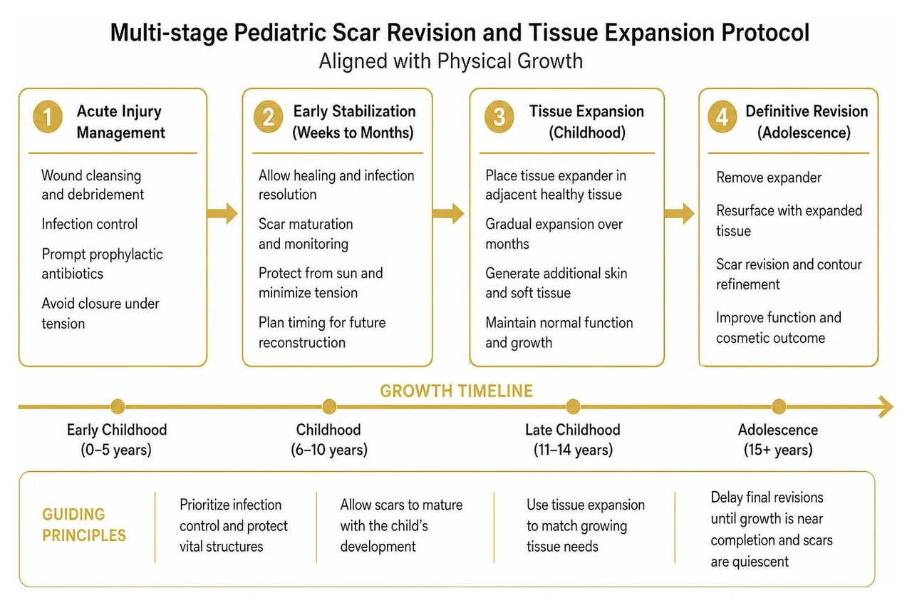
- Delayed Revision Protocols: Because pediatric scar remodeling remains dynamic for several years, reconstructive surgeons generally recommend delaying final scar revisions until physiological maturity or active scar quiescence is reached.
Understanding the full scope of understanding long-term dog bite scarring is essential when projecting the cost of multi-staged revision surgeries required throughout a child's adolescent growth phases.
Systemic Comorbidities and Impaired Scar Maturation
In adult and elderly populations, systemic comorbidities alter inflammatory pathways and delay epithelialization:
- Diabetes Mellitus: Microvascular disease and chronic hyperglycemia impair leukocyte phagocytosis, inhibit capillary proliferation, and reduce fibroblast collagen production, causing prolonged wound open times and fragile, atrophic scars.
- Immunosuppression and Corticosteroid Therapy: Suppressed cell-mediated immunity delays the transition from the inflammatory to the proliferative phase of wound healing, leaving wounds prone to secondary colonization and wound breakdown.
- Peripheral Vascular Disease: Inadequate arterial inflow or venous stasis in the lower extremities deprives healing tissues of oxygen and systemic nutrients, converting simple bite lacerations into chronic, non-healing ulcers that leave thickened, fibrotic margins.
Long-Term Rehabilitation, Scar Revision, and Claim Valuation
Scar tissue never regains 100% of the tensile strength or biological architecture of uninjured skin; under ideal conditions, a healed scar achieves at most 70% to 80% of normal skin tensile strength. Complete scar maturation is a biological process requiring 12 to 18 full months. During this period, immature type III collagen is gradually broken down and replaced with denser, organized type I collagen bundles.
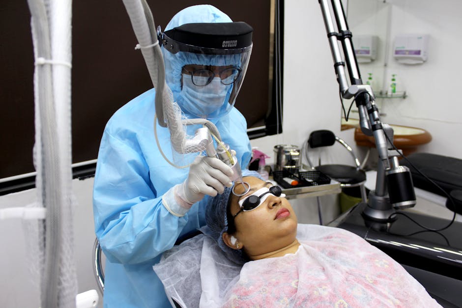
Modern scar rehabilitation requires aggressive, long-term therapies:
- Medical-Grade Silicone Gel and Sheeting: Applied continuously for 3 to 6 months post-epithelialization to maintain tissue hydration and downregulate cytokine signaling that drives fibroblast overproduction.
- Pulsed Dye Laser (PDL) and Fractional CO2 Resurfacing: PDL targets microvascular erythema in early erythematous scars, while fractional ablative CO2 lasers vaporize micro-columns of rigid scar tissue to stimulate smoother, organized collagen realignment.
- Intralesional Corticosteroid Injections: Injections of triamcinolone acetonide directly into active, elevated hypertrophic bands inhibit fibroblast proliferation and flatten dense keloidal tissue.
- Surgical Scar Revision (Z-plasty, W-plasty, or Local Tissue Flaps): Performed after complete scar maturation (typically 12+ months post-injury) to break up straight, contracture-forming scar vectors, reorienting the scar lines into natural relaxed skin tension lines (RSTLs).
Reviewing our comprehensive dog bite injury guide for 2026 helps bite victims understand how these structured clinical interventions interface with personal injury protection and liability insurance coverages.
Assessing Dog Bite Scar Injury Severity for Fair Settlement Compensation
Insurance adjusters frequently push to settle animal attack claims within weeks of the incident. Settling an injury claim before a scar has achieved complete medical stability is one of the most costly errors an injured victim can make.
Evaluating claim value requires establishing:
- Maximum Medical Improvement (MMI): The clinical point at which scar tissue has fully matured and will not improve significantly without invasive surgical revision.
- Future Medical Life-Care Expenses: Comprehensive financial projections accounting for plastic surgery fees, operating room expenses, anesthesia, serial laser treatments, and postoperative medical scar management.
- Permanent Anatomical and Aesthetic Disfigurement: Objective evaluations of scar visibility, pigmentation mismatch, contour depression, and facial asymmetry under various lighting and dynamic facial expressions.
- Loss of Earning Capacity and Vocational Impact: The professional impact of visible facial disfigurement or hand stiffness on public-facing professionals, models, actors, or precision laborers.
Our legal team provides detailed insights into calculating dog bite claim value, leveraging our past insurance-defense background to anticipate how insurance companies attempt to devalue severe scarring claims.
Psychological Trauma and Social Impact of Disfiguring Scars
The true severity of a dog bite scar cannot be measured solely with a millimeter ruler. The psychological damage resulting from sudden animal attacks and visible physical disfigurement often outweighs the acute physical pain.
Bite victims frequently suffer from:
- Post-Traumatic Stress Disorder (PTSD): Clinical studies confirm that up to 24% of severely injured animal bite victims develop chronic PTSD, characterized by sleep disturbances, recurring flashbacks of the attack, hypervigilance, and panic episodes.
- Cynophobia (Severe Fear of Dogs): Debilitating anxiety triggered by barking, neighborhood walks, or visiting friends and family who own pets.
- Pediatric Behavioral Regression: Enuresis (bedwetting), extreme separation anxiety, school avoidance, and social withdrawal in young children following facial trauma.
- Body Dysmorphia and Social Anxiety: Profound self-consciousness, depression, and loss of self-esteem caused by visible facial, neck, or hand scars, forcing individuals to alter their clothing, hairstyle, or daily public interactions.
Under Michigan law, these non-economic injuries represent compensable elements of pain, suffering, mental anguish, and loss of life's daily enjoyments.
Frequently Asked Questions About Dog Bite Scars
What medical factors determine dog bite scar injury severity?
Dog bite scar severity is determined by wound depth, mechanism of trauma (crush, avulsion, or deep puncture), the degree of devitalized tissue, bacterial infection, anatomical location (especially wounds over mobile joints or delicate facial structures), and patient-specific healing factors such as age and glycemic control.
Why should victims wait for full scar maturation before settling an injury claim?
Scar tissue requires 12 to 18 months to undergo complete collagen remodeling and reach Maximum Medical Improvement (MMI). Settling before this biological window closes leaves victims unable to claim compensation for late-stage hypertrophic formation, contractures, or the substantial future costs of laser therapies and surgical scar revisions. Once you sign an insurance release, you cannot reopen the claim.
How does early wound infection worsen long-term scarring?
Infection introduces acute inflammation, bacterial toxins, and localized tissue necrosis. This destructive process breaks down surgical repairs (wound dehiscence), prolongs the healing timeline, and forces the body to lay down excessive, haphazard type III collagen, leading to thick, raised hypertrophic or keloid scars.
Conclusion: Securing Full Value for Permanent Bite Scars
Permanent scars are lasting reminders of a traumatic event. When insurance adjusters evaluate dog bite claims, their primary goal is minimizing exposure by securing a premature release before the true cosmetic and functional severity of the scar becomes permanent.
At Liss, Shapero & Mitnick, our legal team utilizes years of former insurance-defense experience to level the playing field for injury victims across Berkley, Metro Detroit, and throughout Michigan. We know the exact medical metrics, expert plastic surgery evaluations, and psychological assessments required to establish the full, uncompromised value of your case.
Before speaking with an insurance adjuster or signing any settlement release, explore our detailed resource on dog bite scar medical timeline and settlement value drivers, or reach out to Anthony Shapero and the team at Liss, Shapero & Mitnick to ensure your rights and future medical needs are fully protected.
Authored by Anthony Shapero, Managing Partner at Liss, Shapero & Mitnick.
NBK430852.Dhillon J, Hoopes J, Epp T. "Scoping decades of dog evidence: a scoping review of dog bite-related sequelae.." Canadian journal of public health = Revue canadienne de sante publique, 2019. PMCID PMC6964408.Drumright B, Borg B, Rozzelle A, Donoghue L, Shanti C. "Pediatric dog bite outcomes: infections and scars.." Trauma surgery & acute care open, 2020. PMCID PMC7292034.Carlos Altamirano-Arcos, Enrique Chávez-Serna, Israel Espino-Gaucín, Laura Andrade-Delgado, Luciano Nahas-Combina, Rogelio Martínez-Wagner. "Experiencia en el manejo de heridas por mordedura de perro en un hospital de tercer nivel de cirugía plástica y reconstructiva en México." Cirugía y Cirujanos, 2019. DOI 10.24875/ciru.19000678.Kienesberger B, Arneitz C, Wolfschluckner V, Flucher C, Spitzer P, Singer G, Castellani C, Till H, Schalamon J. "Child safety programs for primary school children decrease the injury severity of dog bites.." European journal of pediatrics, 2022. PMCID PMC8821379.Yi‐Shang Yu, Lin‐Yin Wang, Shi‐Wei Yang, Chih‐Hsin Wang. "Choice of Primary Repair in Animal Bite Wound: A Novel Management Strategy." International Wound Journal, 2025. DOI 10.1111/iwj.70761.