
What to Do About a Hematoma After a Dog Bite
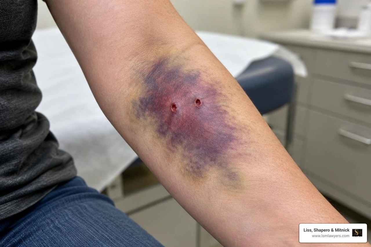
Wash the bite with soap and running water, apply steady pressure if it is bleeding, and cover it with a clean bandage. If a swollen, painful lump or spreading bruise appears, get medical care promptly. Do not try to drain it yourself. Go to the emergency department if the swelling grows quickly, bleeding will not stop, or the area becomes numb, pale, or cold.
A hematoma after a dog bite is blood trapped in tissue. Even a small puncture can hide deeper damage, so the size of the skin wound does not tell the whole story.
I’m Anthony Shapero, managing partner at Liss, Shapero & Mitnick. My years defending insurers before representing injured people shape how I help Michigan clients document injuries such as a hematoma after dog bite trauma.
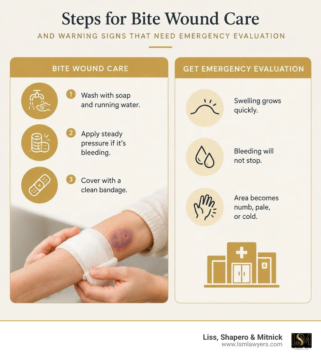
Hematoma after dog bite terms made easy:
Why Dog Bite Appearance Masks Deep Hematomas and Vascular Damage
A common mistake after an animal attack is evaluating the injury solely by surface cuts. When assessing what a dog bite looks like, tiny puncture marks can easily conceal catastrophic underlying trauma.
Canine jaws generate immense crushing force, ranging from 44.96 pounds per square inch (PSI) up to 4,610.74 PSI (310 kPa to 31,790 kPa) depending on the breed. This biting force causes blunt-force trauma, crushing subcutaneous fat, shearing muscle planes, and tearing blood vessels while leaving small skin punctures.
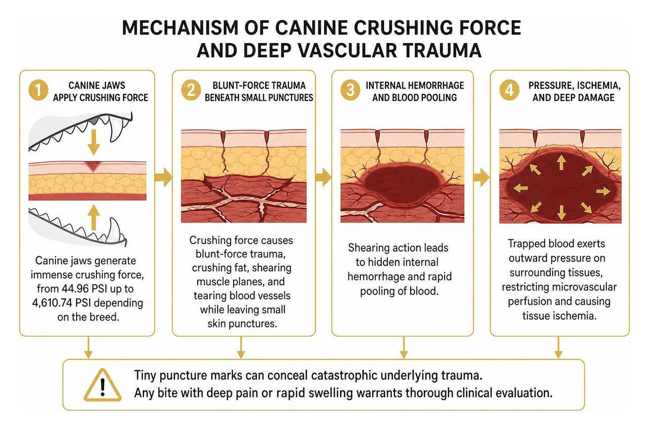
This shearing action leads to hidden internal hemorrhage and rapid pooling of blood. Trapped blood exerts outward pressure on surrounding tissues, restricting microvascular perfusion and causing tissue ischemia. Following established Cleveland Clinic animal bite guidelines, any bite producing deep pain or rapid localized swelling warrants thorough clinical evaluation, regardless of how minor the surface puncture appears.
Anatomical Variations: Extremities, Abdominal Wall, and Scrotal Hematomas
The location of a canine strike dictates the clinical risk profile:
- Upper and Lower Extremities: Extremities account for 80.2% of all dog bite injuries, and 84% of severe bite presentations involve the limbs. When deep canine teeth pierce the forearm or upper arm, they can tear muscular branches or cause direct arterial injury—such as brachial artery occlusion—leading to expanding intramuscular hematomas and limb-threatening ischemia.
- Subungual Regions: Bites crushing the fingertips often produce painful subungual hematomas, where blood pools beneath the nail plate under high pressure, risking permanent nail matrix deformity and osteomyelitis.
- Abdominal Wall: Attacks to the trunk can tear muscular layers without puncturing the peritoneal cavity. A clinical case study on massive abdominal wall hematomas highlights how shearing forces in the flank or iliac fossa can tear inferior epigastric or deep circumflex vessels, accumulating over 500 mL of trapped blood within the oblique musculature.
- Scrotum and Genitalia: The loose, highly vascular tissue of the scrotum allows massive fluid accumulation. An adult scrotal trauma case report documented how canine blunt force fractured the dense tunica albuginea (which requires approximately 110 pounds or 50 kg of force to rupture), causing a massive testicular hematoma and tissue necrosis despite an external laceration measuring just 0.5 cm.
How to Identify and Manage a Hematoma After a Dog Bite
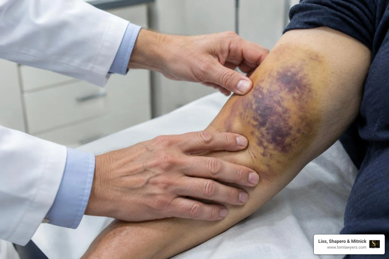
Identifying a hematoma requires physical palpation and advanced diagnostic imaging. Clinicians examine the area for firmness, warmth, and fluctuation—the classic wave-like sensation beneath the skin indicating a localized, unstable fluid collection.
| Characteristic | Simple Post-Traumatic Hematoma | Expanding Vascular Injury / False Aneurysm | Infected Hematoma / Abscess |
|---|---|---|---|
| Onset | Develops over 12–24 hours, then stabilizes | Rapid progression; pulsatile enlargement | Delayed (24–72+ hours post-bite) |
| Palpation | Firm or fluctuant; localized tenderness | Tense, expansile; palpable thrill or bruit | Warm, indurated borders with soft center |
| Skin Changes | Ecchymosis (purple, blue, yellowing) | Pallor, severe tension, shiny skin | Erythema, warmth, red lymphangitic streaking |
| Systemic Signs | None; normal vitals | Tachycardia, hypotension if blood loss is severe | Fever (>100.4°F), chills, leukocytosis |
| Primary Risk | Cosmetic deformity, delayed reabsorption | Acute compartment syndrome, limb loss | Necrotizing soft tissue infection, sepsis |
Warning Signs of a Dangerous Hematoma After a Dog Bite
A stable hematoma gradually stabilizes, but a compromised vessel or invasive infection creates distinct medical emergencies:
- Hard Vascular Signs: Pulsatile bleeding, an actively expanding mass, a palpable thrill, audible bruit, or absent distal pulses.
- Soft Vascular Signs: Diminished pulses, asymmetric coolness, localized nerve deficits, or unexplained hypotension.
- Compartment Syndrome: The "6 Ps"—unbearable pain out of proportion to exam findings, pain on passive muscle stretch, pallor, paresthesia, poikilothermia (coolness), and late pulselessness.
- Invasive Infection / Necrotizing Fasciitis: Severe systemic toxicity, skin crepitus (gas in tissues), purple bullae, and pain spreading rapidly beyond the initial bite margins.
Diagnostic Imaging and Early Surgical Decompression
When physical examination leaves diagnostic doubt, emergency teams employ objective imaging. Point-of-care duplex ultrasound evaluates real-time fluid volume and assesses distal blood flow. For deep wounds or suspected arterial compromise, CT angiography (CTA) maps active vascular extravasation and delineates deep muscular hematomas.
If imaging confirms active arterial bleeding, severe tissue tension, or large fluid collections, surgical evacuation is essential. Surgeons decompress the fascial compartments, evacuate clotted blood, ligate bleeding vessels, and debride devitalized tissue. Primary wound closure is generally contraindicated in contaminated bite wounds with large dead spaces, as suturing traps virulent oral bacteria and accelerates abscess formation.
Medical and Clinical Protocols for Dog Bite Hematomas
Managing dog bite wounds requires a rigorous clinical protocol:
- High-Pressure Irrigation: Copious mechanical flushing using sterile isotonic saline reduces bacterial loads and flushes out foreign debris.
- Conservative Debridement: Removing ragged, non-viable wound edges eliminates necrotic breeding grounds for pathogens.
- Tetanus and Rabies Prophylaxis: A tetanus booster is administered if the patient's last dose was more than 5 years ago. Rabies post-exposure prophylaxis is evaluated immediately if the animal's vaccination status is unknown or unavailable for a 10-day quarantine.
- Injury Staging: Documenting baseline tissue trauma is crucial for both reconstructive planning and evaluating dog bite scar injury severity down the road.
Evidence-Based Antibiotic Prophylaxis and Infection Control
Roughly 3% to 18% of all dog bites become infected, with Pasteurella multocida present in about half of infected wounds. Because canine saliva contains a complex polymicrobial mixture of aerobes (Staphylococcus, Streptococcus) and anaerobes (Fusobacterium, Bacteroides), empiric antibiotic therapy is mandatory for any bite involving a hematoma.
Standard first-line and alternative regimens include:
- First-Line Oral Therapy: Amoxicillin-clavulanate (Augmentin) 875/125 mg orally twice daily for 7 to 10 days provides comprehensive aerobic and anaerobic coverage.
- Alternative Regimens (Penicillin-Allergic Patients): A combination of clindamycin (300 mg four times daily) or metronidazole (500 mg three times daily) PLUS either a fluoroquinolone (ciprofloxacin/levofloxacin), doxycycline (100 mg twice daily), or trimethoprim-sulfamethoxazole (TMP-SMX).
- Inpatient Intravenous Therapy: Ampicillin-sulbactam (Unasyn) 1.5 g to 3.0 g IV every 6 hours for severe, expanding, or surgically evacuated hematomas.
Step-by-Step Patient Guide: At-Home Care and Recovery

Once cleared by an emergency physician or surgeon, recovery focuses on protecting the tissue while the body reabsorbs trapped blood.
How to Treat a Hematoma After a Dog Bite at Home Safely
- Rest and Elevation: Keep the injured limb elevated above heart level for the first 48 to 72 hours to decrease capillary hydrostatic pressure and minimize swelling.
- Cold Therapy Protocol: Apply an ice pack wrapped in a clean, dry cloth for 15 to 20 minutes every 2 to 3 hours during the initial 48 hours. Never apply bare ice directly to damaged skin.
- Clean Dressing Changes: Change non-stick sterile bandages daily, or immediately if the dressing becomes wet, soiled, or loose.
- Prescribed Medications: Complete the entire course of prophylactic antibiotics, even if external symptoms improve quickly.
- Strict Contraindications: Never attempt to drain, needle-aspirate, squeeze, or incise a hematoma at home. Puncturing intact skin introduces external surface bacteria directly into trapped blood, turning a sterile hematoma into an aggressive abscess.
Red Flag Symptoms Requiring Immediate Emergency Care
Return to an emergency department immediately if you notice:
- A sudden spike in fever above 100.4°F (38°C) or systemic chills
- Rapidly expanding redness, purple skin discoloration, or red streaks traveling toward the heart
- Foul-smelling drainage or cloudy pus leaking from puncture points
- Escalating pain that does not respond to prescribed medications
- New or worsening numbness, tingling, or cool, pale skin distal to the injury
Frequently Asked Questions About Dog Bite Hematomas
How long does it take for a hematoma from a dog bite to heal?
Small, uncomplicated subcutaneous hematomas typically resolve within 2 to 4 weeks as macrophages clear cellular debris and break down hemoglobin. Deep intramuscular or abdominal wall hematomas can take 6 to 12 weeks or longer to fully resorb, often leaving behind a firm, temporary scar-tissue knot (induration) as deep muscle fibers remodel.
What is the difference between a dog bite hematoma and an infection?
A simple hematoma is a sterile collection of blood that develops within hours of mechanical trauma, presenting with deep purple bruising, localized firmness, and stable or declining pain. An infection develops later (typically 24 to 72 hours post-injury) and is characterized by increasing warmth, spreading bright-red erythema, purulent discharge, foul odor, and systemic signs such as fever.
Can a dog bite hematoma lead to permanent tissue damage or scarring?
Yes. Unchecked, high-pressure hematomas compress surrounding microvessels, leading to local tissue necrosis, full-thickness skin loss, and severe hypertrophic or keloid scarring. Furthermore, if a hematoma becomes secondarily infected, the resulting tissue destruction can create permanent contour deformities and chronic functional deficits.
Conclusion
A hematoma following a dog attack is more than a bad bruise—it is an indicator of serious blunt-force trauma, torn blood vessels, and high infection risk.
At Liss, Shapero & Mitnick, our legal team draws on decades of former insurance-defense experience to advocate for injured victims across Berkley, Metro Detroit, and throughout Michigan. We know how insurance adjusters attempt to minimize severe internal crush injuries as superficial bruising. If you or a loved one suffered deep tissue trauma, obtaining dog attack legal help ensures your medical bills, surgical costs, and long-term damages are fully covered.
Protect your legal rights from day one by documenting medical timelines and photographs of your wound progression, and reach out to Anthony Shapero and our experienced team for a dedicated case evaluation.