
When a Facial Dog Bite Needs Cosmetic Surgery

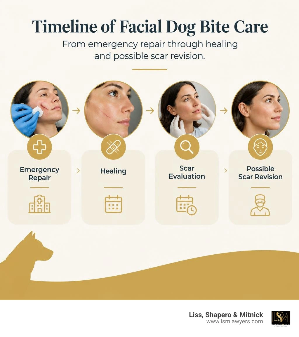
Dog bite cosmetic surgery may be needed when a bite tears away skin or damages the lips, nose, eyelids, nerves, or deeper tissue. A surgeon may repair those structures during emergency care, then treat a lasting scar after the wound has healed. If the bite is on your face, deep, or missing tissue, seek urgent medical care and ask whether a board-certified plastic surgeon should evaluate it.
The first repair matters, but it may not be the last. Scars can tighten or change as they heal, so a final decision about scar revision often comes months later. For Michigan families, that can mean planning for future care as well as the first hospital bill.
I'm Anthony Shapero, a Michigan personal injury lawyer who has represented both insurers and injured people. That experience helps me identify the records and future costs that matter in dog bite cosmetic surgery claims.
Similar topics to dog bite cosmetic surgery:
The Role of Dog Bite Cosmetic Surgery in Facial Tissue Repair
An estimated 45 million dog bites occur across the United States every year, with approximately 800,000 victims requiring professional medical intervention. When an animal strikes, the face is disproportionately targeted—accounting for over 44,000 facial injuries annually. Roughly three out of five facial bite victims are young children whose height puts them directly at eye level with domestic canines.
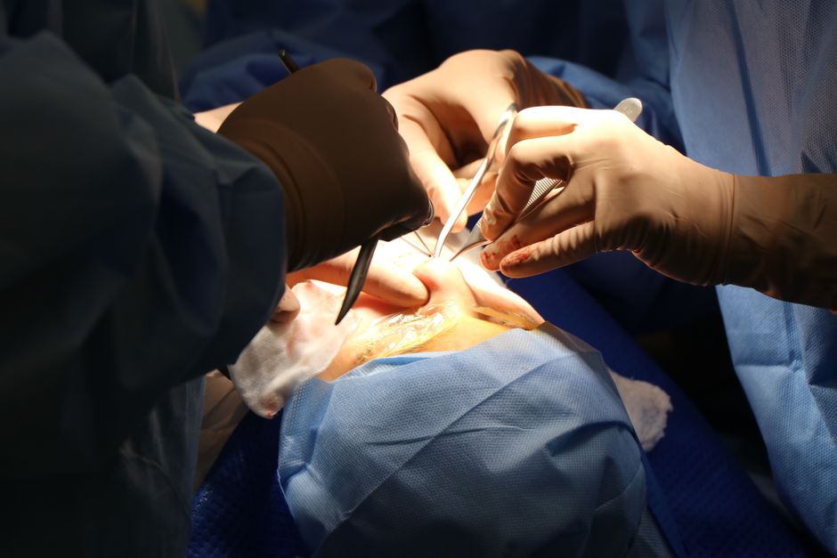
Unlike a clean incision from glass or a surgical scalpel, a canine bite delivers complex, high-energy trauma. When teeth sink into flesh, the animal's powerful jaw exert crushing forces that can exceed 300 to 450 pounds per square inch. When the animal instinctively shakes or pulls its head, severe shearing and avulsion forces tear soft tissue away from underlying muscular and skeletal attachments.
When you are evaluating what a severe dog bite looks like, you are rarely looking at a simple cut; you are looking at devitalized skin, fragmented margins, and exposed subcutaneous structures.
Proper management requires an intimate understanding of Soft Tissue Management in Facial Trauma (Braun & Maricevich, 2017). Facial anatomy is organized into distinct aesthetic subunits—such as the forehead, eyelids, nose, cheeks, lips, and chin. Preserving the delicate boundaries between these units is vital for facial symmetry and emotional expression. When an injury disrupts complex neurovascular networks or tears through cartilage, early evaluation by a board-certified plastic or reconstructive surgeon is essential to prevent permanent, disfiguring deformities.
Mechanisms of Facial Trauma and Tissue Loss
The mechanical trauma of a dog bite typically involves a combination of puncture wounds, deep lacerations, and full-thickness tissue avulsion. The canine teeth create deep, inoculating punctures that drive oral flora into fascial planes, while the carnassial teeth grip and crush tissue margins.
This crushing action compromises microvascular perfusion. Even if torn skin appears physically attached, the underlying capillaries are often thrombosed, rendering the tissue ischemic. Key anatomical landmarks are particularly vulnerable:
- The Vermilion Border: The sharp junction between the red lip and surrounding skin. A misalignment of even one millimeter creates an instantly noticeable cosmetic defect and disrupts the underlying orbicularis oris muscle sphincter.
- Nasal Cartilage: Bites to the nose frequently shear through the delicate alar rims and lower lateral cartilages, leading to structural collapse and airway obstruction if uncorrected.
- Orbital Margins and Eyelids: Trauma near the eyes can sever the canalicular system (tear ducts), damage the levator palpebrae muscles (causing ptosis), or tear the eyelid margin, preventing complete eye closure and exposing the cornea to ulceration.
Why Emergency Room Closure Is Often Insufficient
Emergency departments excel at life-saving stabilization, hemorrhage control, and initial infection prevention. However, standard emergency wound management focuses on rapid linear closure to achieve immediate skin coverage.
In a busy trauma bay, jagged or crushed wound edges may simply be pulled together under high mechanical tension. Suturing ischemic, non-viable wound margins under tension frequently causes:
- Marginal tissue necrosis and wound breakdown.
- Widened, raised, or hypertrophic scars.
- "Trapdoor" deformities, where curved lacerations heal as raised, pincushioned mounds of tissue.
- Distortion of mobile facial structures, pulling the eyelid downward (ectropion) or twisting the corner of the mouth (oral commissure).
When assessing dog bite scar severity, we routinely see that simple linear approximations performed in the ER require comprehensive secondary revision down the road. Plastic surgeons employ meticulous debridement, tissue undermining, tension-relieving deep dermal sutures, and anatomical realignments that emergency physicians are simply not equipped or scheduled to perform.
Surgical Techniques and Timelines Across Reconstructive Stages
Restoring facial form and function after severe canine trauma requires a staged surgical roadmap. Reconstructing the face is not a single event; it is an evolving process that balances immediate infection risks with long-term aesthetic goals.
| Surgical Technique | Primary Indication | Clinical Mechanism | Key Advantages | Primary Limitations |
|---|---|---|---|---|
| Primary Layered Closure | Clean lacerations without significant tissue loss | Precise anatomical realignment using deep, absorbable tension-reducing sutures | Immediate repair, minimal donor morbidity, fast recovery | Unsuitable for wide avulsion defects or severely crushed skin |
| Local Tissue Flaps (e.g., Abbé, Kazanjian) | Full-thickness tissue loss of lips, nose, or cheeks | Mobilization of adjacent tissue with an intact, dedicated blood supply | Excellent match for skin color, texture, and thickness | Creates secondary local scars; requires advanced surgical planning |
| Composite Grafting | Avulsed ear segments, alar rim loss, or salvaged lip tissue | Free transfer of multi-layered tissue (skin, fat, cartilage, or mucosa) | Preserves original anatomical contours without major flap elevation | Highly reliant on recipient bed vascularity; risk of partial necrosis |
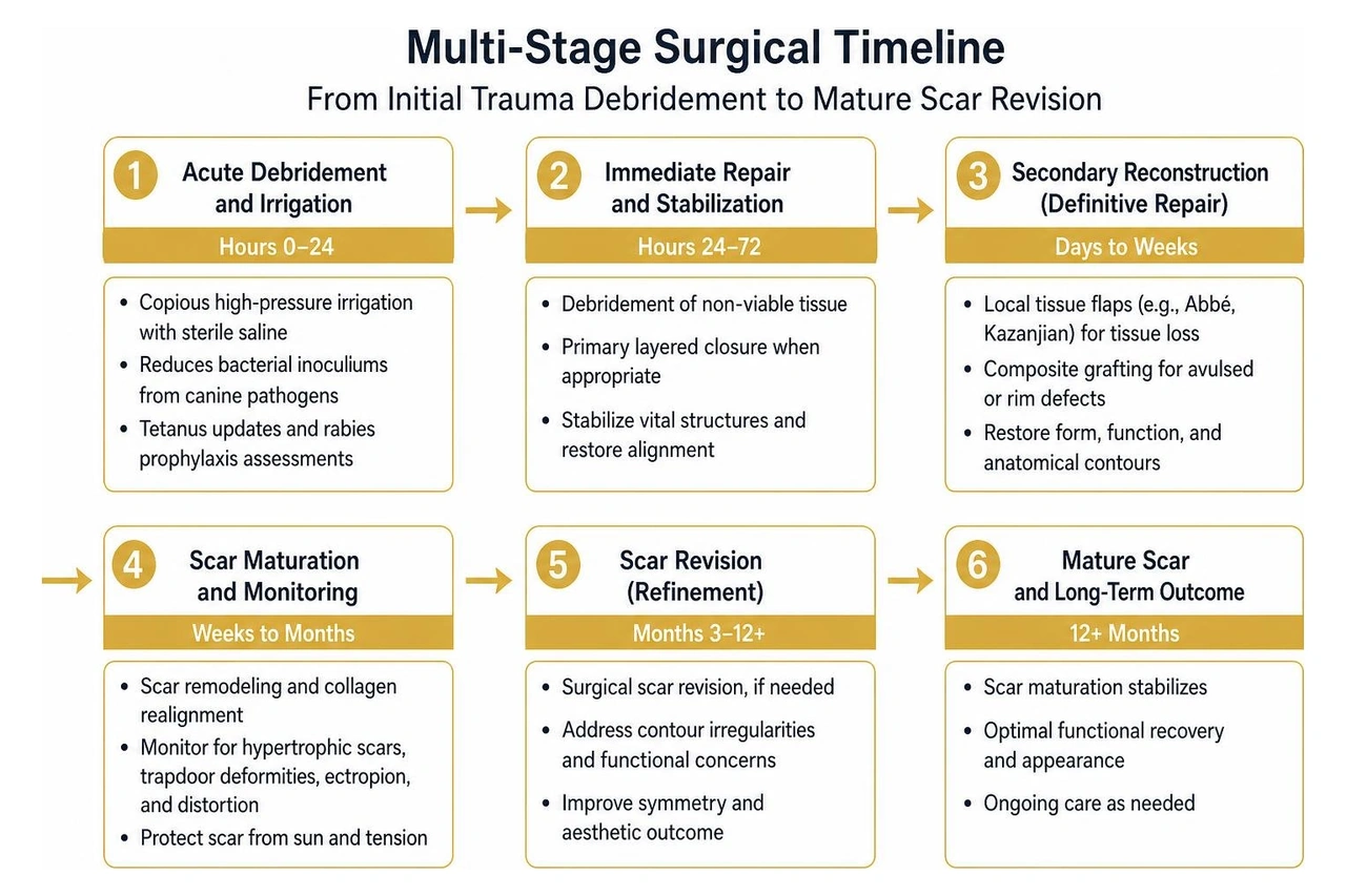
Understanding the medical timelines for dog bite scarring allows patients and legal advocates to anticipate the clinical and financial requirements of long-term recovery.
Immediate Interventions: Debridement and Salvaged Tissue Grafts
The acute phase begins within hours of the attack. Copious high-pressure irrigation with sterile saline is mandatory to reduce bacterial inoculums from canine pathogens like Pasteurella multocida, Staphylococcus, and Capnocytophaga. Tetanus updates and rabies prophylaxis assessments must occur immediately.
Surgeons carefully debride crushed, non-viable margins while conserving every millimeter of healthy tissue. When an attack causes complete tissue avulsion—such as tearing off an earlobe, a portion of the cheek, or a lip segment—any retrieved tissue should be brought to the hospital wrapped in sterile saline gauze and placed on ice (never frozen directly).
In specialized centers, surgeons can clean and defat these salvaged segments to create composite myomucocutaneous or chondrocutaneous autografts (Sheth & Thakur, 2024). If major vascular pedicles remain intact or are repairable under operative microscopy, microvascular replantation can re-establish immediate blood flow, sparing the patient extensive secondary donor-site morbidity.
Complex Tissue Avulsion and Staged Dog Bite Cosmetic Surgery
When tissue is completely lost and cannot be replanted, reconstructive surgeons turn to advanced local and regional flap techniques. The lips present an intricate challenge because they must maintain both oral competence (preventing drooling and allowing normal eating) and symmetrical mobility for speech and expression.
For extensive upper lip defects exceeding 70% tissue loss, advanced surgical approaches combine local advancement and cross-lip techniques. Research on Upper Lip Flap Reconstruction demonstrates that combining a Kazanjian flap (advancing local cheek and mucosal tissue) with a cross-lip Abbé flap (transferring a triangular segment of the lower lip based on the labial artery) successfully restores oral competence and facial symmetry. In staged procedures, the Abbé flap remains connected between the upper and lower lips for roughly two to three weeks to establish microvascular integration before the pedicle is divided in a minor secondary surgery.
For wider facial defects, surgeons may insert subcutaneous tissue expanders—silicone balloons gradually inflated with saline over several months—to stretch adjacent healthy skin, creating matching, hair-bearing skin flaps that seamlessly replace scarred or missing tissue.
Reconstructive Strategies for Highly Melanated Skin
Patients with darker complexions (Fitzpatrick Skin Types IV through VI) face distinct biological challenges during post-traumatic wound healing. In these patients, the cutaneous inflammatory cascade frequently triggers severe post-inflammatory hyperpigmentation (PIH), hypopigmentation, or abnormal fibroproliferative responses.
Achieving a subtle cosmetic outcome requires tailored surgical strategies and diligent long-term treatment for dog bite scars.
Preventing Keloids and Hypertrophic Scarring
Hypertrophic scars and keloids affect approximately 4.5% to 16% of the general population, with a significantly higher prevalence in highly melanated individuals (Sheth & Thakur, 2024). While hypertrophic scars remain confined to the boundaries of the original bite wound, keloids invade surrounding healthy skin, forming raised, pruritic, and painful nodules of dense type-I and type-III collagen (Sheth & Thakur, 2024).
To mitigate these risks, surgeons and dermatologists employ aggressive, multi-modality preventative protocols:
- Early Silicone Gel and Sheeting: Applying medical-grade silicone creates an occlusive, hydrating barrier that normalizes fibroblast signaling and reduces collagen deposition.
- Custom Pressure Therapy: Elastic pressure garments or specialized facial clips exert continuous mechanical pressure, decreasing capillary perfusion to the scar and starving overactive fibroblasts of oxygen.
- Intralesional Corticosteroid Injections: Serial micro-injections of triamcinolone acetonide (Kenalog) directly into developing scars inhibit collagen synthesis, reduce cellular proliferation, and flatten raised tissue.
- Adjuvant 5-Fluorouracil (5-FU): In refractory cases, blending low-dose 5-FU with corticosteroids arrests fibroblast activity without causing localized tissue atrophy or hypopigmentation.
Specialized Incision Planning and Post-Surgical Care
Minimizing abnormal scarring in melanin-rich skin begins with strategic surgical execution:
- Aligning Along Relaxed Skin Tension Lines (RSTLs): Every incision or flap margin must follow natural facial expression lines and wrinkle creases. Perpendicular cuts experience higher mechanical tension, stimulating excessive scar formation.
- Minimal Operative Trauma: Surgeons handle tissue with delicate skin hooks rather than crushing forceps, preserving the fragile dermal-epidermal junction.
- Multi-Layered, Tension-Free Closure: Deep, long-lasting absorbable sutures (such as polydioxanone) anchor the deep dermal and muscular layers, taking 100% of the mechanical tension off the epidermal surface.
- Meticulous Subepidermal Suturing: Non-reactive, fine monofilament sutures (e.g., 6-0 or 7-0 polypropylene) are placed subcuticularly and removed early (within 4 to 5 days) to eliminate "railroad track" cross-hatch marks.
- Strict Ultraviolet (UV) Photoprotection: Exposure to UV radiation accelerates melanin synthesis within healing scars, causing permanent dark discoloration. Daily broad-spectrum physical sunscreen (zinc oxide/titanium dioxide) is non-negotiable for at least 12 to 18 months post-injury.
Advanced Scar Revision and Non-Surgical Refinements
Even with flawless initial surgery, complex bite lacerations frequently heal with irregular contours, structural shadowing, or webbed scar contractures that restrict facial mobility.
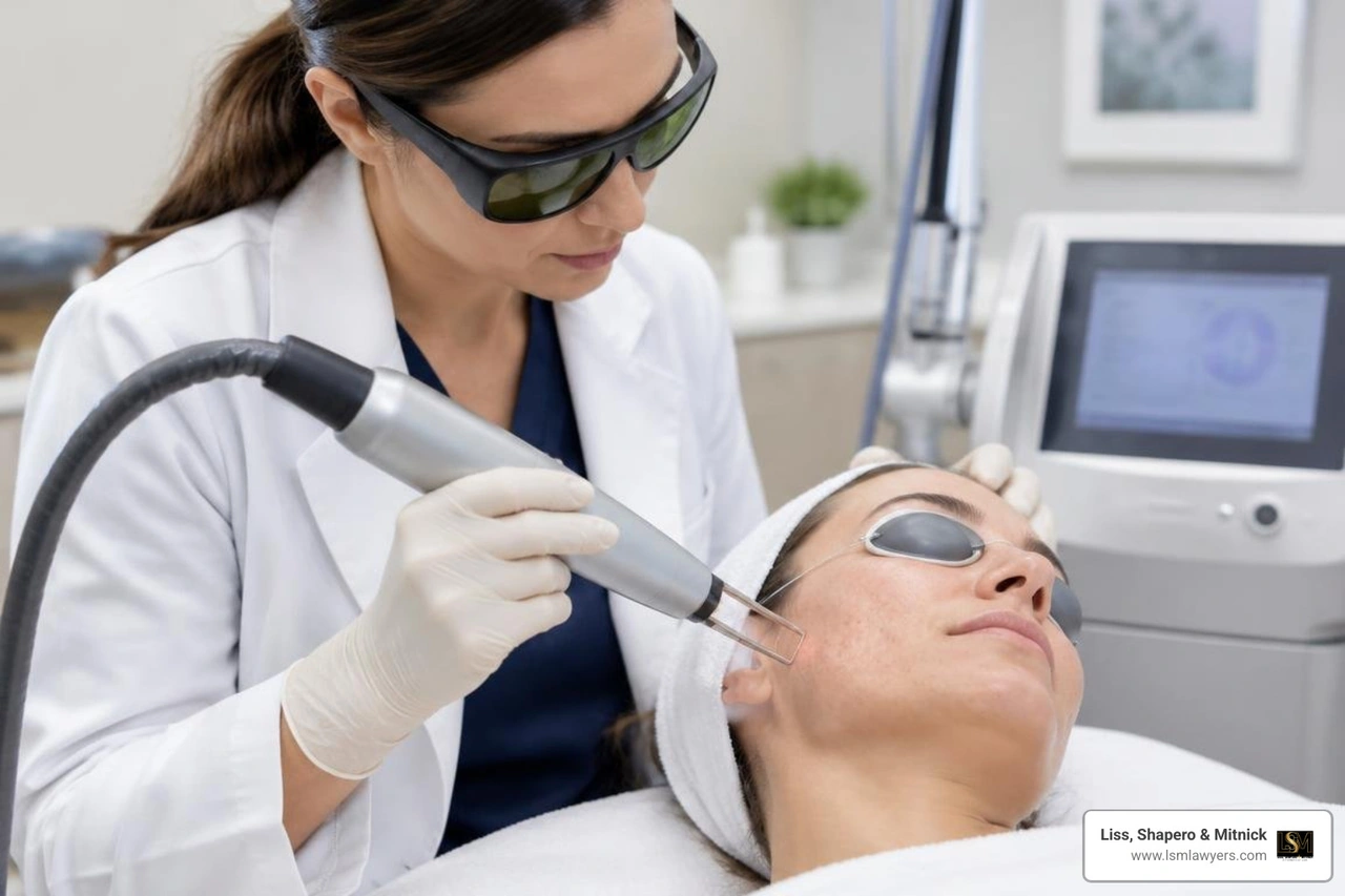
Once initial wound healing is complete, secondary scar revision refines tissue texture, levels uneven margins, and repositions conspicuous lines to blend into natural facial shadows, as detailed in our comprehensive dog bite injury guide.
Optimal Timing for Secondary Dog Bite Cosmetic Surgery and Revision
Patience is a crucial clinical requirement. Immature scar tissue goes through prolonged metabolic phases:
- Proliferative Phase (Weeks 1 to 6): Scars appear red, raised, hard, and hypervascular.
- Early Remodeling Phase (Months 2 to 6): Collagen cross-linking increases, and tissue begins to soften.
- Mature Remodeling Phase (Months 6 to 12+): Vascularity recedes, and the scar reaches its final tensile strength (approximately 80% of uninjured skin).
Revising a scar too early, while active cellular remodeling is underway, can cause severe inflammation and poor surgical outcomes. Surgeons generally wait at least 6 to 12 months for full scar maturation before undertaking definitive geometric scar revisions:
- Z-Plasty: A transposition technique that changes the directional axis of a scar, breaking up a straight line into a zig-zag pattern that aligns with natural skin folds while relieving tight, restricting tissue bands.
- W-Plasty: Resecting scar edges into interlocking triangular interdigitations to disrupt light reflection across flat surfaces, making long, linear scars far less noticeable.
- Geometric Broken-Line Closure (GBLC): Excising the scar and creating irregular, interlocking geometric shapes (squares, triangles, rectangles) that confuse the human eye, preventing it from detecting a continuous surgical line.
Non-Invasive Modalities: Lasers, Botulinum Toxin, and Microneedling
Modern cosmetic revision heavily incorporates non-surgical technologies that optimize dermal remodeling without further scalpel incisions:
- Pulsed-Dye Laser (PDL - 595 nm): Selectively targets microvascular hemoglobin in early, erythematous scars, collapsing abnormal capillary beds and removing the bright red or purple discoloration.
- Fractional Ablative and Non-Ablative Resurfacing (CO2 and Erbium:YAG): Creates microscopic thermal treatment zones in the dermis, vaporizing dense, disorganized collagen and stimulating the production of smooth, healthy new extracellular matrix.
- Botulinum Toxin A (Botox) Injections: Botulinum Toxin Helps Facial Scars Heal Better by temporarily paralyzing underlying facial expression muscles around a healing wound or revision site. Eliminating repetitive muscular pull across the wound edge minimizes tension, leading to significantly narrower, flatter, and more discreet scars.
- Radiofrequency (RF) Microneedling: Delivers controlled thermal energy into the deep reticular dermis to break up dense fibrotic tethering and smooth surface irregularities.
- Hyaluronic Acid Dermal Fillers and Fat Grafting: Restores lost subcutaneous volume beneath depressed, atrophic bite scars, lifting the indented tissue flush with the surrounding skin.
Psychosocial Recovery and Managing Medical Costs
The emotional aftermath of a violent facial attack can be as devastating as the physical wounds. Facial disfigurement strips away a person's sense of self, often precipitating severe post-traumatic stress disorder (PTSD), social anxiety, severe depression, and cynophobia (the pathological fear of dogs).
Children are especially vulnerable. A visible facial scar can trigger bullying, social withdrawal, academic regression, and lasting developmental distress. Cosmetic surgery is not a vanity project; it is an indispensable medical bridge to psychological healing and social reintegration.
These complex, staged treatments carry substantial financial burdens:
- Emergency room trauma fees and operative room debridement: $5,000 to $25,000+
- Plastic surgical reconstruction and microvascular grafting: $10,000 to $50,000+
- Secondary scar revisions (Z-plasty, flap thinning): $4,000 to $15,000 per stage
- Non-invasive laser therapy series, microneedling, and Botox: $3,000 to $10,000+
- Psychological counseling and specialized trauma therapy (EMDR): $2,500 to $10,000+
Health insurers routinely attempt to deny coverage for secondary revisions, laser resurfacing, and dermal structural adjustments, labeling these critical restorative procedures as "purely cosmetic" or "elective."
At Liss, Shapero & Mitnick, our legal team routinely handles these disputes. Because I previously represented insurance companies, I know the exact defense playbooks adjusters use to downplay the severity of facial scarring and discount future surgical estimates.
Securing the full dog bite claim value requires working with board-certified plastic surgeons who can provide comprehensive future-care cost projections. Under Michigan’s strict liability statute (MCL 287.351), dog owners are held legally responsible for bite injuries sustained by unprovoked victims on public property or lawfully on private property. We ensure that settlements and verdicts account not just for immediate emergency room bills, but for every stage of reconstructive surgery, laser revision, and psychological support required over a lifetime.
Frequently Asked Questions About Facial Bite Repair
When should a plastic surgeon be consulted after an animal attack?
A plastic surgeon should ideally evaluate any facial dog bite, full-thickness skin avulsion, or deep wound crossing cosmetic borders within the first 6 to 24 hours. Early specialist intervention ensures precise debridement, anatomical preservation of margins (like the vermilion border), and strategic primary closure that dramatically reduces the need for complex revisions later on.
How long does complete facial scar maturation take?
Complete facial scar maturation takes between 12 and 18 months. During the first three months, scars often appear red, firm, and slightly raised due to hypervascularity and active collagen deposition. Over the following year, the scar gradually softens, flattens, and lightens. Definitive surgical revisions, such as Z-plasty or dermabrasion, are typically scheduled after the six-month mark to ensure the tissue has stabilized.
What first-aid steps preserve tissue before reconstructive surgery?
Immediately apply direct, firm pressure with a clean cloth or sterile gauze to control bleeding. Gently irrigate the wound with clean water or saline—do not use harsh chemicals like hydrogen peroxide or alcohol, which damage exposed cellular structures. If a piece of skin, lip, or ear has been torn off, wrap it in a sterile, saline-moistened gauze, place it inside a sealed plastic bag, and set that bag on crushed ice. Do not submerge the tissue directly in water or let it contact ice directly, as freezing causes tissue necrosis.
Conclusion
Facial dog bites are deeply traumatic, life-altering injuries that require sophisticated, staged medical management. From the initial emergency debridement and composite tissue grafting to complex flap advancements and laser scar revisions, restoring your appearance and facial function demands patience, clinical precision, and specialized surgical care.
Navigating physical recovery while managing rising medical bills and insurance disputes can easily overwhelm any family. At Liss, Shapero & Mitnick, we leverage our deep knowledge of insurance-defense tactics to level the playing field for injury victims across Berkley, Metro Detroit, and all of Michigan. We ensure that negligent dog owners and their insurance carriers are held accountable for the full scope of your past and future care.
If you or your child suffered a severe facial bite and requires surgical reconstruction, you do not have to face the recovery process alone. Reach out to our team for experienced legal representation for Michigan dog bite injuries, and let us fight for the resources you need for a complete physical, emotional, and financial recovery.
— Anthony Shapero, Partner at Liss, Shapero & Mitnick
Medical References
Sheth R, Thakur M. "The Surgical Repair of an Avulsion Dog Bite and Scar Revision in Highly Melanated Skin: A Case Report.." Cureus, 2024. PMCID PMC11668903.Braun TL, Maricevich RS. "Soft Tissue Management in Facial Trauma.." Seminars in plastic surgery, 2017. PMCID PMC5423789.